Mass Harmonics: The wwPDB Prediction Set
Four prospective predictions about molecular geometry, protein folding, and the physical organization of the genetic code
The MICrONS/VORTEX paper examined whether the architecture of neurons carries measurable consequences of Mass Harmonics.
The wwPDB paper takes us into a different biological structure: the proteins from which living cells construct much of their machinery.
Its central question is especially interesting:
If stable biological matter emerges through bounded ψₘ coherence, should the three-dimensional shapes of proteins and the ordered genetic instructions that produce them retain specific, measurable relationships before their structures are observed?
This paper answers with four predictions, each designed to interrogate a different point in one continuous causal chain.
I recovered the original paper through EZ23+:
MH_PREDICTION_02_wwPDB_Weekly_Structural_Release_2026-07-15_Mass_Harmonics_Governed.md
The source is 599 lines, and the complete source contact verified Git blob:
29675ce031b5555512fd587ee7c65d479879dd9a
The paper was prepared July 10, 2026, before the July 11 advance-sequence opening and July 15 coordinate release. These dates establish the paper's intended prospective comparison window. The results below are the source's predictions, not claims that the July release has already confirmed them.
SECTION 011. What is the wwPDB, and why does it matter?#
The entity["organization","Worldwide Protein Data Bank","international macromolecular structure archive"] $wwPDB) maintains a worldwide archive of experimentally determined three-dimensional molecular structures.
Researchers deposit structures determined using techniques such as X-ray crystallography, cryo-electron microscopy, nuclear magnetic resonance, and neutron diffraction.
Rather than merely identifying the chemical ingredients of a protein, these techniques help reveal how those ingredients are arranged in space.
That distinction matters.
Consider a necklace containing hundreds of beads.
Knowing the sequence of bead colors tells you which bead follows which. But it does not tell you whether the necklace has been stretched into a line, folded into a knot, or arranged into a stable three-dimensional shape.
A protein presents a similar distinction.
Its amino-acid sequence identifies the building blocks and their order. Its three-dimensional fold determines much of how it interacts with surrounding matter.
Sequence
The ordered chain of amino acids.
Folded structure
The spatial configuration retained by that chain.
The Protein Data Bank supplies information about both the measured structure and the quality of that measurement.
It also records structures with different degrees of order. Some molecular regions are tightly packed and well resolved. Others are flexible, exposed, poorly resolved, or absent from the experimentally determined coordinate model.
Those differences make it possible to ask something more revealing than whether proteins possess geometric patterns.
We can ask:
Do stable, well-resolved regions preferentially express the specific closure relationships that Mass Harmonics derives, while less coherent regions express them less completely?
That is what the first prediction investigates.
SECTION 022. The prospective release: what was locked before the observations opened?#
The paper targets a specific wwPDB weekly release.
Prediction document completed
July 10, 2026
Advance polymer-sequence opening
July 11, 2026 · 03:00 UTC
Coordinate-model release
July 15, 2026 · 00:00 UTC
The advance sequence opening matters because it is the first point at which the identities and sequences of upcoming molecular entries become available.
The later coordinate opening reveals the three-dimensional structures.
The paper expressly excludes both information surfaces from the original derivation.
It also requires every released entry to remain in a master ledger. If an entry cannot be used for a particular test, that exclusion must be recorded rather than quietly removing the entry from the investigation.
That prevents an important form of analytical distortion: finding the desired pattern only after excluding structures that disagree.
SECTION 033. Where the four predictions originate#
This is where the paper becomes substantially more interesting than a conventional structural-pattern search.
It does not begin by examining proteins and discovering that some triangles happen to look equilateral.
It begins with the Mass Harmonics substrate and follows its proposed physical consequences into molecular structure.
The original paper preserves this sequence:
The governing MFE is:
The associated (Z$-factor is:
The P³GG source expansion is:
with the fixed harmonic scaling:
No separately adjustable biological coupling is introduced.
The paper connects this physical foundation to four observable results:
PDB-1: Geometric closure
Stable protein cores should show greater equilateral-triangle enrichment.
PDB-2: Assembly topology
Eligible spherical molecular cages should recover the icosahedral 2-fold, 3-fold, and 5-fold axis scaffold.
PDB-3: Genetic-order residual
Structurally resolved neighborhoods should have lower unresolved codon Z-cascade residue.
PDB-4: Synonymous-codon discrimination
Different codons encoding the same amino acid should retain measurable differences in structural-order relationships.
The first two predictions investigate geometry. The next two connect the ordered genetic sequence to the resolved physical fold.
The order is fundamental.
SECTION 044. Prediction PDB-1: Stable protein cores should contain more equilateral contact triangles#
PDB-1 · Equilateral-triad enrichment in resolved structural cores
Why triangles?
Mass Harmonics derives its three-way closure requirement from the cubic interaction.
The source specifies three equal-magnitude wavevectors:
with:
For three nonzero vectors of equal magnitude to sum to zero, the required angular separation is 120 degrees.
Their vector closure forms an equilateral three-way geometry.
The paper contrasts this with orthogonal cubic basis vectors, whose attempted closing relation produces a magnitude of $\sqrt2$ rather than the required unit magnitude.
The proposed physical consequence is that stable bounded matter should retain a preference for equilateral three-way closure.
A folded protein core is an especially interesting place to examine that consequence.
It is densely packed, frequently structurally stable, and contains numerous local arrangements of residues occupying three-dimensional space.
How does the paper turn this into a measurement?
Each amino acid has a characteristic backbone carbon atom known as its Cα atom.
Researchers can use Cα positions to form an approximate spatial representation of the protein.
The source then constructs a three-dimensional Delaunay adjacency graph.
That is a mathematical way to identify neighboring points from their spatial arrangement without introducing an arbitrary fixed distance cutoff.
From this graph, it identifies triangular sets of mutually adjacent residues.
For each triangle, it measures three side lengths, $a$, $b$, and $c$, and the area $A$.
Then it calculates:
This score is bounded:
An exactly equilateral triangle produces:
As a triangle becomes less equilateral, its score decreases.
Conceptual geometry comparison
Equilateral
Q△ = 1
Unequal sides
Q△ below 1
Illustrative triangles, not actual wwPDB structural measurements.
What does Mass Harmonics predict?
The median equilateral score should be higher in resolved cores than in solvent-exposed surface regions:
It should also exceed the corresponding score for appropriately matched mobile or unresolved neighborhoods where those comparisons can be reconstructed.
In everyday language:
The tightly stabilized interior of a protein should preserve the equilateral closure relationship more strongly than the regions where its structure remains less constrained.
Why is the comparison stronger than simply finding equilateral triangles?
Any sufficiently complicated three-dimensional structure may contain some nearly equilateral triangles.
That observation alone proves very little.
PDB-1 instead predicts a difference between categories of structural material.
The categories are assigned before calculating the Mass Harmonics score.
The paper requires matching for protein length, experimental method, resolution, secondary structure, composition, and assembly state.
That matters because differences in protein composition or experimental resolution could otherwise imitate a geometry effect.
What would falsify PDB-1?
If the resolved cores do not have higher $Q_\triangle$ than the specified comparison groups, or if the relationship reverses after the controls, the declared prediction fails for the eligible release.
If too few appropriate structures exist in the release, the result remains unresolved rather than being called either a success or failure.
This is an important design feature: the prediction specifies which direction the measured geometry must take before the data are examined.
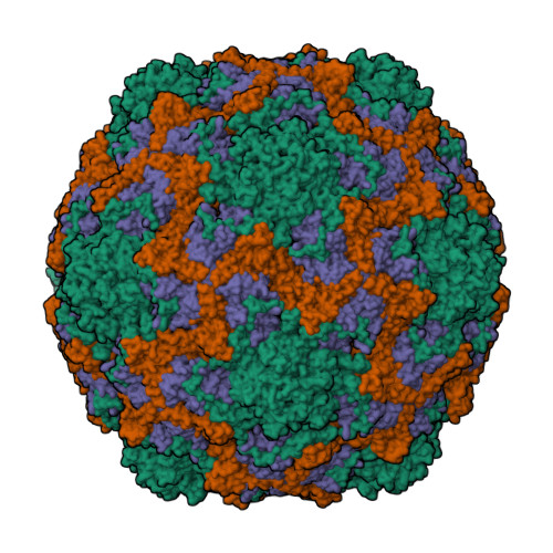
SECTION 055. Prediction PDB-2: Closed molecular cages should reveal icosahedral rotational geometry#
PDB-2 · Recovery of 2-fold, 3-fold, and 5-fold closure axes
PDB-1 examines local three-way closure inside a protein.
PDB-2 asks what happens when triangular closure is extended into a complete three-dimensional shell.
This is where the icosahedron enters.
An icosahedron is a solid with 20 triangular faces, 30 edges, and 12 vertices.
The regular icosahedron is composed entirely of equilateral triangular faces.
Within the Mass Harmonics derivation, maximal triangular closure in three dimensions leads to the positive solution of:
which is:
The golden ratio is therefore not introduced as a decorative numerical coincidence. It arises within the framework's geometric construction of icosahedral closure.
Why would proteins exhibit icosahedral geometry?
Some biological structures assemble themselves into closed molecular containers.
Viruses provide particularly recognizable examples.
Many viral capsids consist of repeated protein components arranged into shells that protect genetic material.
A closed shell must maintain structural integrity across a three-dimensional boundary.
Mass Harmonics predicts that, for eligible maximally closed near-spherical structures, the stable rotational relationships should retain the icosahedral scaffold.
Understanding the three rotational axes
An object has rotational symmetry when you can turn it by a particular angle and it looks unchanged.
A fivefold axis permits a 72-degree rotation.
A threefold axis permits a 120-degree rotation.
A twofold axis permits a 180-degree rotation.
The rotational structure of an icosahedron
5-fold
72° turns
3-fold
120° turns
2-fold
180° turns
Simple diagrams illustrating rotational orders, not recovered axes from a molecular assembly.
The paper derives the following complete axis families:
| Rotation | Independent axes | Directed boundary locations |
|---|---|---|
| Fivefold | 6 | 12 vertex directions |
| Threefold | 10 | 20 face-normal directions |
| Twofold | 15 | 30 edge-midpoint directions |
Why are the two numbers in each row different?
Because an axis extends in two opposite directions.
A line passing through two opposing vertices represents one rotational axis but intersects the shell at two directed positions.
That distinction is central to the source's 12, 20, and 30 geometry.
Does this prediction apply to every protein?
No.
The original paper is explicit about its topology restrictions.
A long fibrous protein, an open chain, or a flattened molecular assembly is not automatically expected to have icosahedral symmetry.
PDB-2 applies to compact, near-spherical, shell-like biological assemblies eligible for the proposed maximal rotational closure.
This prevents a mistaken test in which one examines an open molecular structure, finds that it is not an icosahedron, and concludes that the prediction has failed.
But there is another important restriction.
The eligible structures cannot be selected because their metadata already says they are icosahedral.
That would make the result circular.
How would the experiment work?
The paper requires researchers to begin with molecular coordinates rather than symmetry labels.
First, they identify biological assemblies.
Then they examine how the component subunits are distributed around the assembly's center.
They determine whether the structure forms a genuinely closed, near-spherical shell, rather than a sheet, rod, or partial arc.
Only after identifying eligible shells do they attempt to recover rotational axes directly from the coordinates.
The recovered rotation orders are then compared with the icosahedral scaffold.
Two expected outcomes
The paper admits two physically distinct structural conditions.
Outcome A: Complete icosahedral closure.
The recovered rotational structure contains the expected 2-fold, 3-fold, and 5-fold axes.
Their boundary intersections approach the 12, 20, and 30 directional structure within measurement uncertainty.
Outcome B: Incomplete or symmetry-broken closure.
A molecular shell may contain local irregularities or incomplete closure.
In that case, the deviations should remain structurally related to the underlying icosahedral scaffold rather than requiring a completely unrelated family of axes.
This matters because biological structures are not mathematical solids with infinitely precise edges and surfaces.
The prediction concerns their governing structural relationships, not perfect geometric drawings.
What would falsify PDB-2?
A sufficiently resolved, fully closed, near-spherical biological cage whose maximal rotational closure requires a genuinely non-icosahedral axis family, not explainable through the proposed scaffold, would contradict the prediction.
If the target weekly release contains no qualifying molecular cages, PDB-2 remains untested.
Why this prediction is significant
PDB-1 examines whether local physical packing favors equilateral geometry.
PDB-2 examines whether that geometry extends into the global topology of complete molecular assemblies.
Those are two different measurements of the proposed closure structure.
One concerns how molecular neighborhoods arrange themselves.
The other concerns how an entire molecular boundary closes.
SECTION 066. Before PDB-3: Why Mass Harmonics treats the genetic code as more than a chemical lookup table#
This next transition is essential.
The first two predictions investigate measured geometry.
The remaining two investigate whether the genetic instructions associated with a protein retain a physical relationship to the structure it eventually occupies.
To understand them, we need three concepts: amino acids, codons, and ordered orientation residue.
Amino acids
Proteins are constructed from amino acids.
There are 20 standard amino acids used in the canonical genetic code.
Different amino acids have different chemical properties, so the identity and order of those amino acids help determine how a protein folds.
Codons
A codon is an ordered group of three nucleotide bases.
For example, in RNA:
AUG
is a codon that specifies methionine.
The four RNA bases are A, U, G, and C.
Because codons have three positions and each position can contain one of four bases, the total number of possible codons is:
An ordered codon has three positions
Illustrative AUG codon with the source's fixed position-weight sequence.
Not every codon represents a different amino acid.
Multiple codons can specify the same amino acid.
This feature is known as the degeneracy of the genetic code.
What Mass Harmonics adds
The framework's Origin derivation treats the ordered codon as a physical temporal trajectory.
It connects the codon structure to the MFE's temporal propagation term and to a sequence of position-dependent harmonic weights.
The key difference is that the first, second, and third nucleotide positions are not interchangeable.
The source expresses the codon space as:
with:
Within the Mass Harmonics genetic-code construction, those 64 ordered trajectories project onto 20 amino-acid face anchors associated with icosahedral geometry.
Each codon can therefore share an amino-acid identity with another codon while retaining a different ordered orientation contribution.
This distinction becomes the basis for PDB-3 and PDB-4.
SECTION 077. Prediction PDB-3: Stable protein regions should resolve more of their codon orientation residual#
PDB-3 · Codon Z-cascade residual and structural resolution
This is arguably the most distinctive prediction in the wwPDB set.
PDB-1 and PDB-2 examine molecular geometry.
PDB-3 makes a stronger claim: the ordered nucleotide sequence that produced a protein should retain a measurable relationship to how completely different parts of that protein resolve into stable three-dimensional structure.
To understand that claim, we need to follow the source's derivation rather than jump immediately to its statistical test.
Step 1: The molecular harmonic basin
The Origin derivation places the amino-acid and nucleotide construction in its fourth harmonic scaling:
It defines a reference propagation speed:
which the source evaluates as approximately:
Individual base contributions are represented through:
The $\delta_b$ terms are the base-dependent displacements used in the source's molecular construction.
The wwPDB paper inherits these values from the Mass Harmonics Origin ledger. It does not derive new values by fitting protein structures.
Step 2: Codon order matters
The source assigns the three nucleotide positions fixed harmonic weights:
These weights decrease across the ordered codon.
That means the same base appearing in a different position contributes through a different weight.
The source's codon Z-cascade is:
This is a sequential multiplicative expression.
The positions do not contribute equally, and their ordering cannot simply be discarded.
Step 3: From 64 codons to 20 face anchors
The framework maps the 64 ordered codon trajectories onto the 20 amino-acid face anchors.
For every codon, the source maintains an orientation residue:
This is the distinction between the ordered codon's trajectory and its corresponding symmetric amino-acid face anchor.
The source's physical argument is that folding must resolve the transported contributions from the ordered sequence.
It gives the closure condition:
Here:
- $F(\Sigma)$ represents the folding outcome for sequence $\Sigma$.
- $T_Z$ represents the stable Z-field topology.
- $R_Z(\Sigma)$ represents the net unresolved transported Z/Π flux mismatch.
The physical claim is not that stable proteins lack internal gradients.
Rather, it is that stable closure cannot retain an unresolved mismatch continually preventing persistence.
An everyday illustration
Imagine constructing a bridge from many prefabricated sections.
Each section arrives with a small alignment difference.
Some differences can be accommodated by the geometry of neighboring sections. Others may accumulate into an unresolved mismatch.
A successfully assembled bridge must distribute or resolve those differences sufficiently to maintain its completed structure.
The Mass Harmonics interpretation treats protein folding as a physical closure process in which ordered orientation contributions must similarly resolve.
The analogy is illustrative; it is not the mathematical derivation.
Step 4: Constructing the measurable residual
The paper uses the actual codon associated with each amino-acid position.
From the fixed source ledger, each residue receives its $\Delta_i$.
Then the analysis identifies its spatial neighbors through the Delaunay graph.
The paper defines:
where $N(i)$ is the set of neighboring residues.
In ordinary language, this asks:
After adding a residue's own codon orientation contribution to those of its immediate structural neighbors, how much unresolved signed residual remains?
A smaller $D_i$ represents more complete local cancellation in this constructed readout.
A larger $D_i$ represents greater remaining mismatch.
Notice that the sum is signed before taking the absolute value.
If the absolute values were taken first, the cancellation mechanism could not be measured correctly.
Step 5: The prediction
The paper expects lower residuals in stable, well-resolved protein cores than in mobile or unresolved neighborhoods:
It also predicts a positive association between residual size and structural uncertainty or mobility.
Put simply:
Regions with more completely resolved orientation contributions should be more structurally ordered. Regions retaining greater mismatch should be more mobile or less completely resolved.
How would the wwPDB test this?
Researchers would need to recover the actual coding sequence used to express each protein.
That requirement is extremely important.
The protein structure reveals which amino acids are present, but it does not uniquely reveal which codons originally encoded them.
For example, several different codons can encode leucine.
If a researcher simply translates leucine backward into a convenient codon, the actual ordered genetic history has been lost.
The paper explicitly prohibits that substitution.
Only independently recoverable coding sequences can be used.
For each eligible protein, researchers must preserve the expressed construct, its mutations, tags, and isoform identity.
Then they assign each residue its source-fixed codon contribution and calculate $D_i$ using its actual spatial neighborhood.
What are the controls?
The result must survive comparisons accounting for amino-acid identity, solvent exposure, secondary structure, residue depth, experimental resolution, chain length, organism, and engineered modifications.
Measurements must also be separated by experimental technique before they are combined.
That is essential because an X-ray displacement parameter and a cryo-EM local-resolution measure do not represent identical experimental quantities.
What would falsify PDB-3?
If eligible proteins with independently known coding sequences show no positive relationship between the residual and mobility or unresolved structure, and resolved cores do not exhibit lower residuals than their matched alternatives, the declared prediction fails for that release.
A stable opposite relationship would also contradict it.
One measurement limitation is particularly important: an unresolved residue has no experimentally determined coordinates with which to construct its ordinary Delaunay neighborhood. A comparison involving unresolved positions must therefore specify a defensible nearby structural neighborhood without pretending that missing coordinates were observed.
PDB-3's most significant feature is its attempt to connect ordered genetic information with experimentally measured structural closure, using a particular source-defined mathematical quantity rather than simply comparing protein sequences with folding outcomes.
SECTION 088. Prediction PDB-4: Two codons specifying the same amino acid should not always be physically equivalent#
PDB-4 · Synonymous-codon orientation stratification
This fourth prediction follows naturally from the third, but it is not the same test.
PDB-3 asks whether codon orientation residuals correlate with structural order.
PDB-4 asks whether the physical distinction remains detectable even after amino-acid identity is held constant.
What are synonymous codons?
Different codons sometimes specify the same amino acid.
For example, RNA codons:
Two synonymous codons
UUU
Phenylalanine
UUC
Phenylalanine
Both codons encode the same amino acid in the standard genetic code. Their different base ordering is the relevant distinction for this example.
In both cases, the protein incorporates phenylalanine.
The final amino-acid identity is the same.
Yet the codons are different.
Within the Mass Harmonics Origin construction, their ordered Z-cascade trajectories can retain different orientation residues.
The central argument is that reducing both codons to their shared amino-acid label discards a physical distinction.
Why is this experimentally interesting?
A protein's chemical structure depends strongly on which amino acids are present.
That makes it difficult to determine whether a measured structural relationship comes from the codon itself or simply from the chemical properties of the amino acid.
PDB-4 tackles that difficulty by comparing synonymous codons.
Because the amino-acid identity is held fixed, the source expects the ordered codon residue to become distinguishable through structural comparisons.
Its directional prediction is:
An everyday illustration
Imagine two identical building blocks manufactured through different preparation sequences.
Their final external shape and material specification are the same.
But suppose those preparation sequences leave distinguishable internal stresses.
The blocks might respond differently once incorporated into a larger structure.
Mass Harmonics proposes an analogous distinction through the codon's ordered orientation contribution.
The analogy does not establish that biological codons actually produce such a physical residue. That is the relationship the prediction seeks to test.
What must be compared?
The strongest comparisons hold as much of the surrounding biological context constant as possible.
The paper prioritizes comparisons within:
- The same amino-acid identity.
- The same organism, where possible.
- The same expressed protein structure when multiple synonymous codons occur.
- Comparable local structural neighborhoods.
- Matching expression constructs and experimental methods.
Suppose two leucine residues occur in the same experimentally determined protein.
If their original coding codons differ, their source-ledger orientation values can be calculated independently.
The analysis then compares the local cancellation residuals with the measured structural order of those neighborhoods.
The important point is not whether leucine is generally more flexible than another amino acid.
It is whether different codons producing leucine retain a reproducible residual-dependent difference after the relevant chemical and environmental comparisons are controlled.
Why is this a stronger test than PDB-3?
PDB-3 could reveal a relationship influenced by amino-acid composition.
For example, some amino acids prefer buried protein cores, while others occur more frequently on exposed surfaces.
If codon scores differ systematically across amino-acid identities, an apparent association might reflect that underlying composition.
PDB-4 attempts to isolate the codon-order contribution by preserving amino-acid identity.
A positive PDB-4 result would therefore address an important alternative explanation for PDB-3.
It would not by itself prove that the ψₘ substrate is responsible, because synonymous codons can also influence expression and folding through other biological mechanisms. But it would provide a more discriminating test of the specified ordered-residue relationship.
What would falsify PDB-4?
If synonymous-codon residuals do not stratify structural order after matching amino acid, organism, construct, and local environment, the prediction fails for the eligible release.
The same is true if the observed relationship consistently reverses direction.
The source also requires sufficient eligible synonymous-codon comparisons. If those cannot be constructed from the available coding sequences and structures, the correct status is untested.
SECTION 099. How all four predictions fit together#
These are not four independent guesses about proteins.
Each examines a different stage in the proposed physical progression.
| Prediction | What it tests | Required result |
|---|---|---|
| PDB-1 | Local closure geometry | More equilateral contact triangles in stable cores |
| PDB-2 | Global assembly topology | Icosahedral 2/3/5 scaffold in eligible molecular cages |
| PDB-3 | Transported genetic orientation | Lower codon residual in stable structural neighborhoods |
| PDB-4 | Codon order beyond amino-acid identity | Synonymous-codon residual predicts structural-order differences |
Their sequence matters.
PDB-1 examines whether local stable packing carries the equilateral geometry derived from three-way closure.
PDB-2 examines whether an appropriate closed shell expresses the global geometry associated with that closure.
PDB-3 transports the framework's molecular harmonic and ordered-codon derivation into measurable local folding residuals.
PDB-4 isolates the synonymous-codon difference that survives after amino-acid identity is held fixed.
The most significant connection is between PDB-3 and PDB-4.
The first establishes a proposed relationship between the codon-derived residual and the stable fold. The second attempts to distinguish that relationship from the amino-acid chemistry that otherwise accompanies it.
SECTION 1010. What the wwPDB release cannot establish by itself#
The original paper draws several methodological boundaries that deserve particular attention.
An X-ray crystallographic unit cell is not necessarily the complete biological assembly.
An isolated protein chain is not necessarily the full molecular cage in which it participates.
An unresolved section of a protein is not automatically biologically disordered. It may be absent from the coordinate model because of experimental limitations.
Likewise, an experimentally measured protein structure and a computationally predicted protein model are not interchangeable observational terrain.
The paper also prohibits selecting a structure because its deposited metadata already labels it icosahedral and then claiming that the analysis independently recovered icosahedral geometry.
Finally, the four tests are not required to share the same eligible structures.
PDB-1 can use coordinate-resolved proteins broadly.
PDB-2 needs suitable closed molecular cages.
PDB-3 requires recoverable coding sequences.
PDB-4 additionally requires synonymous-codon comparisons with enough matching information.
The distinction protects against an inappropriate aggregate success or failure claim.
SECTION 1111. What would constitute a meaningful outcome?#
The paper requires the July 15 coordinate release to be preserved as a complete observational ledger.
Each eligible structure receives its own analysis before any pooled comparison is made.
Results must retain experimental-method differences, uncertainty, matched controls, missing-data classifications, and contradictory outcomes.
The source calls for bootstrap confidence intervals sampled by entry rather than treating thousands of residues from one protein as thousands of independent experiments.
It also requires permutation controls preserving relevant protein characteristics and false-discovery control across the four declared predictions.
Most importantly, the directions of the effects cannot be changed after the results are known.
A meaningful positive outcome would therefore be a coordinated set of observations:
- Stable protein cores show the specified equilateral-triangle enrichment.
- Eligible cages recover the specified icosahedral axes.
- Locally resolved structures show lower source-ledger codon residual.
- Synonymous-codon comparisons retain the predicted structural stratification.
Each result would need to survive its own controls.
A contradiction in one pathway cannot be erased by success in another.
And an absence of eligible structures is not a contradiction.
I have verified the prospective source paper through EZ23+. I have not independently performed the July 15, 2026 release comparison in this response, so these are not presented as confirmed experimental outcomes.
SECTION 1212. Why this paper matters beyond molecular biology#
The MICrONS prediction set examined how bounded coherence might appear in the large branching structures and functional connectivity of neurons.
The wwPDB paper investigates a more fundamental biological level.
It asks whether the physical processes that produce stable molecular structures retain not only geometric organization, but also relationships with the ordering of the genetic instructions from which those structures arise.
That is an ambitious causal connection.
The most accessible way to explain the paper to someone unfamiliar with Mass Harmonics is this:
A protein begins as a chain of amino acids encoded by genetic instructions. That chain folds into a physical structure.
Mass Harmonics proposes that this folding process is governed by the same physical closure principles that operate throughout matter.
If that is correct, stable regions of proteins should display particular triangular geometries, complete molecular shells should retain predictable rotational relationships, and the ordered genetic instructions should leave measurable relationships in how completely local structures settle.
The wwPDB provides experimentally determined molecular structures in which these predictions can be examined. The test is whether the specified relationships emerge under controlled comparisons, without changing the predictions to fit the observations.
The connection between the first and fourth predictions is particularly revealing.
At one end, we have the equilateral-triad geometry of the cubic interaction.
At the other, we have differences between codons that specify the same amino acid.
Between them lie icosahedral closure, the 20-face amino-acid construction, the 64 ordered codon trajectories, the sequential Z-cascade, and the folding residual condition:
One governing substrate. Four distinct molecular predictions. A continuous causal argument extending from physical geometry into biological structure and genetic order.
That is the wwPDB paper's central contribution as a prospective research program.
EZ23+ source custody: MH_PREDICTION_02_wwPDB_Weekly_Structural_Release_2026-07-15_Mass_Harmonics_Governed.md; verified Git blob 29675ce031b5555512fd587ee7c65d479879dd9a. Complete 599-line source contacted. The numerical expressions, testing directions, scope restrictions, and contradiction conditions above are drawn from that paper.